Our five component oral check includes our external, oral soft tissue, occlusion, periodontal, and endodontic exams. This post will cover the periodontal component, with each of the other components being discussed in its own article.
View all articles in our educational series.
Periodontal Status
The fourth component of the oral exam is periodontal status. Gum disease is common in horses, and is one of the most painful dental conditions horses can have. It is a common misconception that this only happens in older horses. However, young horses can have gum disease as well, which is why oral exams in young horses is so important.
Examples of equine periodontal disease
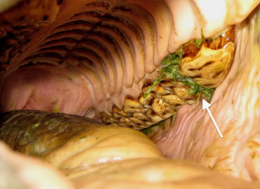
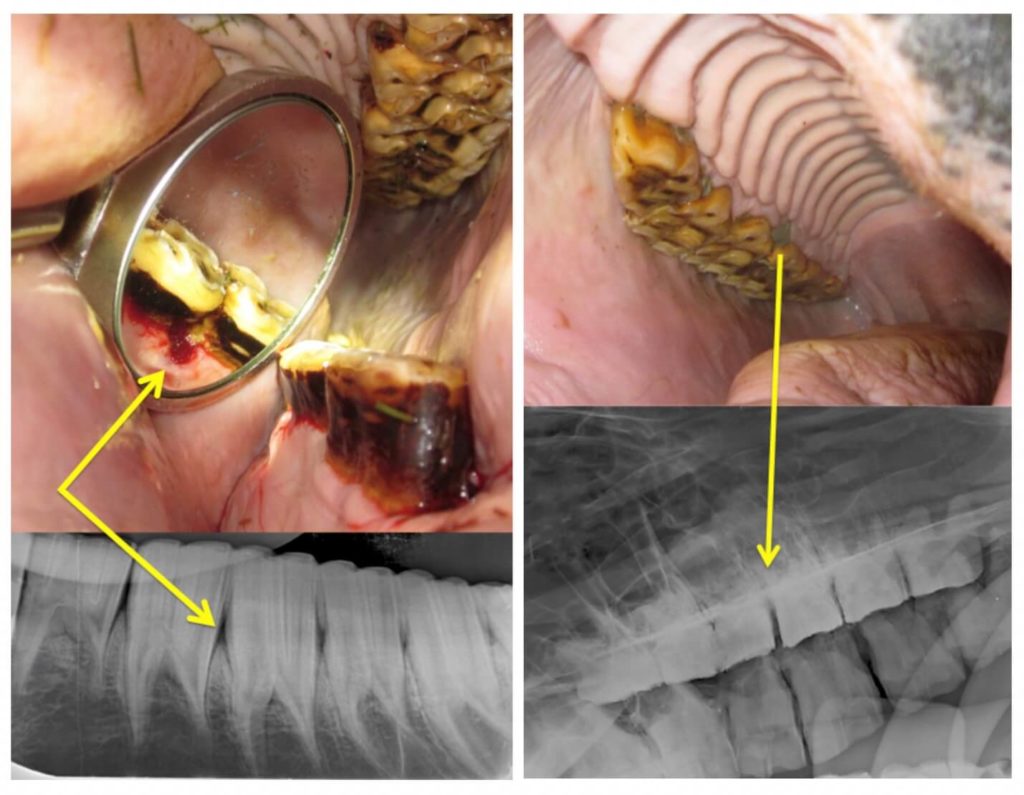
Horses get gum disease when they get feed trapped in between their cheek teeth, which should be tightly packed together. Here is an example of an older horse with a diastema (space with feed packing) in between his first two cheek teeth.
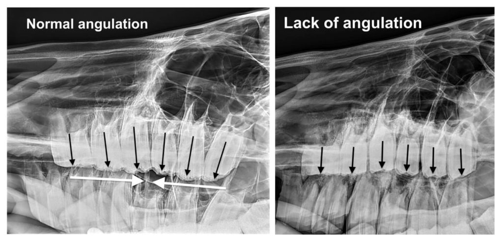
Some horses get these spaces in between their teeth because of POOR ANATOMY. The x-ray on the left is from a horse with normal cheek teeth angulation, which cause the teeth to pack very tightly together in the mouth.
You’ll notice that the horse on the right has teeth that are nearly parallel to one another, which has allowed spaces to form in between all of her teeth.
How do we treat gum disease in horses? There are several ways to approach it.
In all cases, we float the teeth to address high spots that create wedges to further drive the teeth apart. In some cases we perform a periodontal debridement, using a water-pick type device to remove all of the feed from the space.
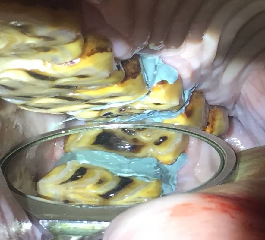
If the space is conducive to it, we will pack impression material as you see in this picture here. This helps improve oral soft tissue comfort and allows the gums to be less irritated. This is not a permanent solution and may need to periodically be repeated.

In some early cases of gum disease, where there is feed trapping and gingival recession but not a large diastema (space) we have a newer technique called occlusal relief cutting that we have had very good success with.
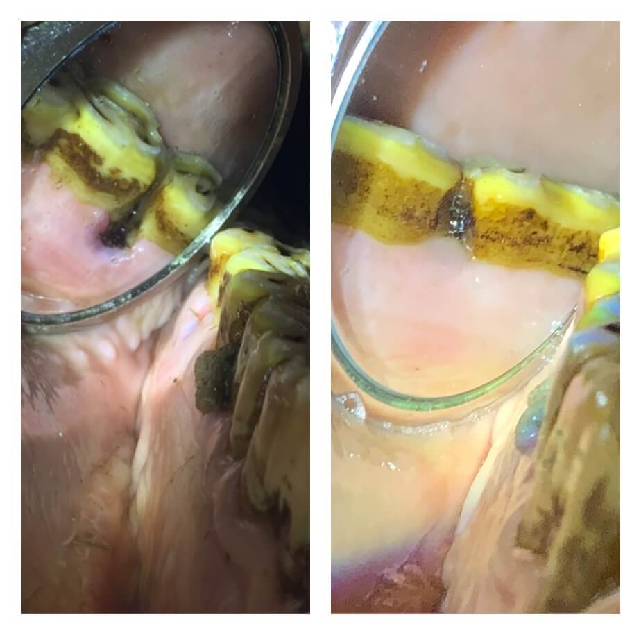
Here is an example of a 17 year old gelding that had a periodontal pocket that we found on his oral exam. There was 8 mm pocket depth (white arrow) with gingival recession. We make a small 3 mm groove (red arrows) at the chewing surface where the two neighboring teeth meet. This allows food to cycle through at the tooth surface, instead of getting trapped at the gumline and in many cases, allows periodontal pocket to completely heal.
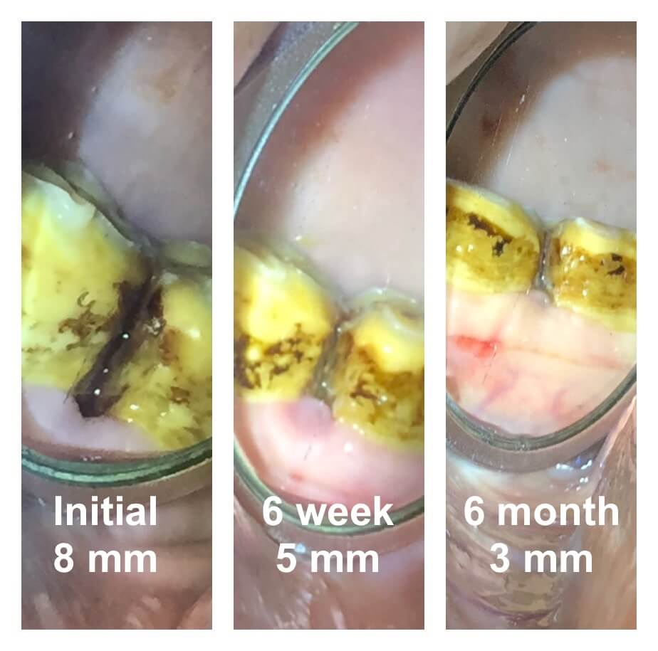
This horse had nearly resolved the periodontal pocket by his 6 week recheck. You’ll notice how the little “scalloped” edge of gingival recession has filled in with more normal gum. Also note how the grey/black discoloration has resolved where the two teeth meet — that is because the food is no longer stagnating in this area.
On the right was 6 months later, and his gums look even better, with only 3 mm depth, which is normal.
Here is another success story with an occlusal relief cut. This horse was completely resolved when we floated his teeth one year later. The important thing about this procedure is to recognize that these horses have anatomical predispositions to developing pocketing in these areas.
Sometimes the occlusal “groove” may have to be repeated again if they start pocketing feed again.
Radiographs are important in a periodontal evaluation because we need to look for bone loss to understand how advanced the disease is.
Gum disease follows a similar pathway to humans — gingival recession (which is the only reversible stage) followed by bone loss, which is permanent. When we find periodontal disease on our oral exam we will often recommend x-rays so that we can come up with the best management plan for your horse.
End stage periodontal disease always ends up in early tooth loss, so an “ounce of prevention is worth a pound of cure!” (Thanks, Benjamin Franklin!)