Our five component oral check includes our external, oral soft tissue, occlusion, periodontal, and endodontic exams. This post will cover occlusion, with each of the other components being discussed in its own article.
View all articles in our educational series.
What is occlusion?
The third component of our oral exam is occlusion. Occlusion means examining how the teeth are aligned in the skull. Deviance from normal can result in malocclusions, which can cause problems for the horse if not addressed.
Malocclusions can be classified in two primary ways—one relates to how the horse was born (skeletal malocclusions), and the other relates to individual teeth and their relationship with other teeth (dental malocclusions).
Examples of occlusion
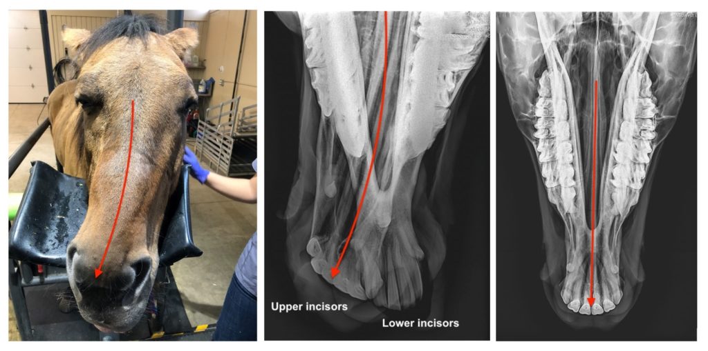
We’ll start with an example of a skeletal malocclusion, which relate to how the horse was born. You’ll see the horse on the left has a very crooked skull—this horse has a wry nose, which is a congenital defect that causes crookedness in the bones of the head. This will affect dental balance, sometimes in a very significant way.
When you look at this horse’s radiographs compared to a normal horse, you will see that his incisors do not line up at all! These horses amazingly can often figure out how to eat, but many require more frequent floats to keep their teeth maintained.
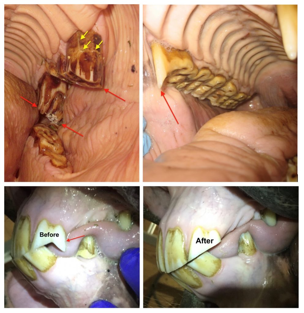
These horses have examples of dental malocclusions. This can happen when teeth are worn, missing, fractured or misaligned, causing the tooth opposite to become overlong as the tooth continues to erupt.
The red arrows show teeth that are overlong due to missing teeth opposing them (upper left picture), or misalignment of teeth due to an overbite (upper right), and lack of alignment of the incisors (bottom pictures before and after floating).
The horse in the upper left picture also has a tooth fracture (yellow arrows) possibly caused by his excessive tooth length.
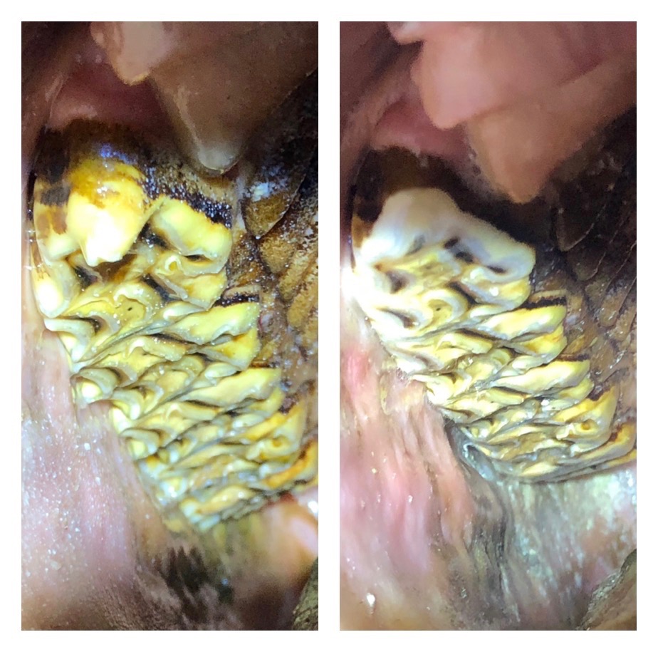
Here is a before and after picture of a horse with a large hook on the first cheek tooth. You’ll notice after floating, we were able to round the front part of the tooth, which will help in bridling comfort.
Here is a before and after picture of a horse with a tooth that was overlong due to a missing tooth in the lower jaw. We were able to remove most of the excess in height, but not all.
When we are floating malocclusions we are always careful to continue monitoring with our dental mirrors to make sure we aren’t getting too close to the sensitive part of the tooth.
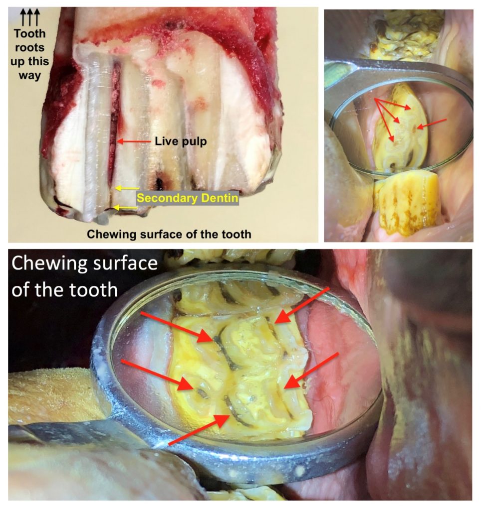
These pictures show a tooth that has been sectioned to show the pulp canal of the tooth. Cheek teeth in horses actually have 5-7 of these canals in each tooth!
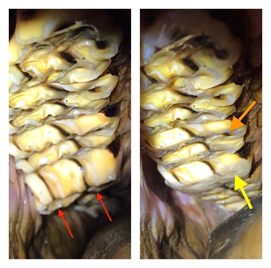
The pulp is the blood and nerve supply of the tooth and needs to stay protected for the tooth to stay alive. Since horses have teeth that continually erupt into the mouth, the live pulp is constantly laying down secondary dentin, which is the dark brown staining material shown by the red arrows in the two mirror pictures.
When we are floating teeth that are too tall, we need to reduce the chewing surface, but must do so carefully to avoid pulp damage or exposure. We monitor the color of the pulp, and stop when it reaches a light brown or tan color as this means we are getting close to the sensitive pulp.
Do you see the change in color in the secondary dentin in the picture on the upper left as it gets close to the live pulp?